“If not for longer throat for Facebook monetization and content money.
Why would NKUBI and his wife be posting their child online, knowing fully well that people will say funny things about her.
They should know that whatever they post online is no longer their business but people’s business to talk or say what ever they want.
If they don’t want people to talk about their baby, then they shouldn’t be posting her online.” ~ A Facebook user known as CRUISE DADDY reveals concerning the new name internet gave to NKUBI’s baby.
👨👩👧 Family Fashion Goals: Stylish Denim Vibes from Ridex Fashion Blog
This stunning family portrait captured by Ridex Fashion Blog radiates warmth, love, and style. Dressed in matching denim outfits, the family showcases a perfect blend of coordinated fashion and natural elegance. Dad rocks a sleeveless white shirt and baggy jeans paired with bold accessories, while mom stuns in a ripped denim jumpsuit and sleek high ponytail. The adorable daughter steals the spotlight in her cute denim overalls and pink sneakers, exuding charm and confidence.
👗 Family Matching Outfits – A Rising Trend
Matching denim is making a strong comeback in family fashion. It not only highlights unity but also creates timeless photos that celebrate togetherness in style. This family proves that with the right outfit, even casual looks can become runway-worthy.
📸 Perfect Look for Family Photoshoots
Whether you’re planning a birthday shoot, holiday card, or just want to capture a memory, this style is a great inspiration. Simple, bold, and full of personality — denim-on-denim is here to stay!
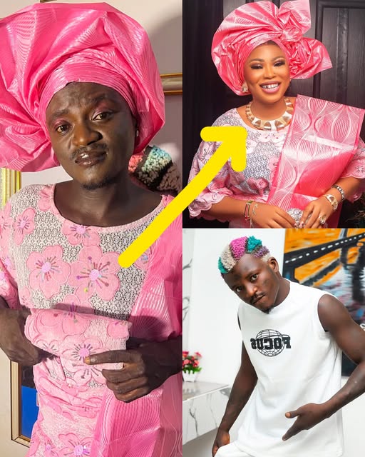
In this impressive photo, the little gentleman wears a traditional Yoruba outfit – a pure white agbada, embroidered with intricate motifs and perfectly combined with a fila hat and trendy black glasses. The boy exudes a confident demeanor, a standard “fashionista” style that makes everyone admire.
👔 Traditional fashion style combined with modernity The outfit not only retains its cultural identity but is also enhanced with modern accessories such as watches and sunglasses, showing creativity in the way it is coordinated. This is proof that traditional fashion can completely become outstanding and trendy.
🔥 Children’s fashion trends 2025 The photo is a typical example of modern children’s fashion trends – where personality, culture and confidence are smoothly combined. It is predicted that in 2025,
the “mini gentlemen” style with a traditional blend will continue to dominate events and festivals.
“Look Down on Me Then, Look Up to Me Now!” — Peller Reflects
“My classmates used to laugh at me in secondary school because I wasn’t book-smart. They didn’t rate me one bit. But fast-forward to today — I’m the biggest streamer in Nigeria, and now they’ll do anything for my attention.
While they were cramming books, I was busy plotting my future — how to make my papa proud.
There was even one guy, Seyi, who used to hide his answers during exams so I wouldn’t copy.
Guess who FaceTimed me recently, screaming that he saw me on TV and begged to visit me and Jarvis in Lekki? I told him, ‘Pay gate fee first.’”
Moral Lesson: Never write anyone off — the same people you mock today might become the ones you pay to see tomorrow.
NDOLO NEWSStella peko et Bojiko sont les heureux parents d’un petit garçon
Félicitations au couple de web comédiens qui accueille leur premier enfant ensemble. Une nouvelle qui console énormément après tout ce par quoi le coupe a dû passer au cours de cette année (accident, maladie, etc ).
#Peupah_Zouzoua Sinon eux jouaient les couples comme ça pourtant eux faisaient vrai vrai hein!?
En tsoukaa la mère et l’enfant se portent bien par la grâce de Dieu. Félicitations à bojiko qui a courageusement supporté les nerfs de la grossesse d’une fille bafang, courte de surcroît ! Teguè interdit, l’envol sera ayop ayop
OWEH NEWS Le père de la docta Agos du paradis a déjà validé Ks Bloom devant nos yeux
Mais qui est Kamga pour recevoir les salutations aussi distinguées du père d’ Indira Baboke?? C’est la question qu’il faut se poser ce matin !
Car lors de la soutenance spéciale de dalida, Samson était présent, dans la même zone que les personnes proches de famille, à l’instar de Samuel Eto’o, tam sir et autres.
#Peupah_Zouzoua Laisson affaire d’humilité, lui quiENT pour recevoir une poignée du papa de la secrétaire du paradis ? Oui c’est un artsite, oui il est respectueux, mais jusqu’à courber la tête et les yeux comme si il avait honte de kokochose ? En tsoukaaa Kamga, tu vas nous dire ce que tu cherches dans le taxi chaque jour au Cameroun! Kooo j’étais avec une fille tout allait bien mais sa famille a dit que c’était pas le moment… HUMMMM !!! Tegue interdit, l’envol sera lancé mais l’oiseau a vu